Learn From Dr Tahseen Riaz

Blog

Stay updated with expert insights, helpful tips, and the latest advancements in orthopedic care to keep your bones, joints, and overall mobility in the best shape.


Blog Page

Some up and coming trends are healthcare consolidation for independent healthcare centers that see a cut in unforeseen payouts with a growth of independent practices.
dr-tahseen-Bone-Fracture.jpeg

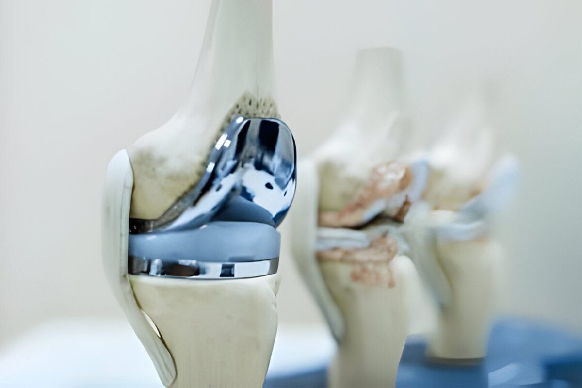
Fractures, commonly called broken bones, can present in various forms and necessitate prompt and precise management. Depending on the severity of the injury, the recovery period for a fracture can range from a few weeks to several months. It is essential to note that mild bone fractures often heal spontaneously, whereas severe fractures may require surgical intervention, immobilization, and the implementation of physiotherapy at various stages of recovery.


What Causes a Bone Fracture?

Bone fractures typically result from traumatic incidents, including automobile accidents, falls from height, and slips leading to awkward landings. The underlying cause of a fracture is generally evident. Moreover, stress fractures can arise from repetitive physical activities, manifesting as hairline fractures in weight-bearing bones, particularly the tibia in the lower leg. These fractures are frequently observed among athletes.

What Are the Types of Bone Fractures?

Bone fractures can be categorized into several distinct types:

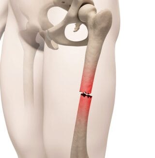
1. Complete Fractures:

Characterized by the bone breaking into two or more separate fragments. These fractures can be further classified by their orientation—transverse, oblique, or longitudinal—and may also present as spiral or winding fractures.

2. Open Fractures (Compound Fractures):

These are complete breaks where the bone pierces the skin, resulting in a higher risk of infection. Such injuries require immediate medical attention.

3. Closed Fractures (Simple Fractures):

In this type, the integrity of the skin remains intact, and the bone does not protrude.

4. Incomplete Fractures:

These involve a crack in the bone without complete separation into two parts, with hairline fractures being a common example.

5. Displaced Fractures:

These complete fractures involve misaligned bone fragments, which often necessitate surgical intervention to realign for proper healing.

6. Comminuted Fractures:

A fracture in which the bone is shattered into several pieces, demands a complex surgical approach.

7. Greenstick Fractures:

These involve fractures that do not extend completely through the bone, which are primarily encountered in children due to the flexibility of their bones.

8. Buckle Fractures:

A specific type of greenstick fracture is where the bone bends without breaking completely.

9. Compression Fractures:

These occur when a bone collapses due to excessive pressure, frequently observed in individuals with degenerative spinal conditions.

10. Avulsion Fractures:

These occur when an injury causes significant tearing of muscle and ligament, resulting in a bone fracture.

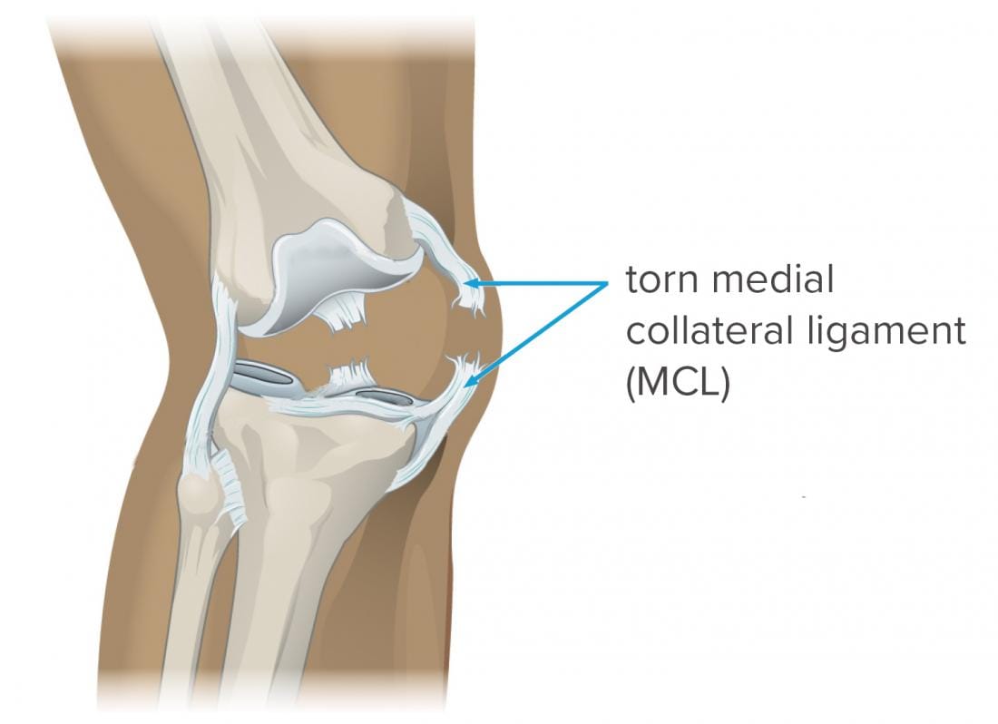
It is important to acknowledge that while dislocations are not classified as fractures, they frequently occur concurrently with fractures in the event of a severe injury.

What Are the Symptoms of a Bone Fracture?

Individuals with bone fractures typically experience acute pain, and dizziness, and may exhibit nausea. The affected limb may be immobile and unable to bear weight. Other symptoms encompass a grinding sensation, bruising, swelling, tenderness, discolouration, and, in the case of open fractures, bleeding. Misalignment of the limb is a conspicuous indicator, particularly in instances of complete fractures.

 Who Is at Risk of Bone Fractures?

Although all individuals are susceptible to bone fractures, certain populations are at heightened risk:

Older Adults:

Many individuals over the age of 50 demonstrate symptoms of osteoporosis, leading to thinner and more fragile bones. Approximately 50% of women and 20% of men sustain a fracture after the age of 50.

Post-Menopausal Women:

The reduction of estrogen levels significantly increases the risk of developing osteoporosis.

Athletes and Dancers:

Participants in contact sports such as hockey and football are at risk of various fracture types, including open fractures. In addition, non-contact sports may lead to stress fractures, while extreme sports such as horseback riding, cycling, and skiing pose risks for severe injuries. These individuals must wear appropriate protective gear.

Individuals with Chronic Health Conditions:

Those with chronic illnesses, including diabetes, osteomyelitis, rheumatoid arthritis, ongoing infections, and cancers that metastasize to the bones, are more vulnerable to fractures.

Individuals With Poor Lifestyle Choices:

A diet deficient in essential vitamins and minerals elevates the risk of osteoporosis. Regular physical activity is essential, and a diverse exercise routine is critical in preventing stress fractures. Maintaining proper nutrition, engaging in consistent exercise, and ensuring adequate sunlight exposure for vitamin D synthesis are fundamental preventive measures.

Children:

Due to their developing bones and greater flexibility, children are particularly susceptible to greenstick and buckle fractures.

 What to Do If Someone Has a Bone Fracture

In cases of suspected skull fractures, compound fractures, or other serious injuries, it is imperative to seek emergency medical assistance without delay. Do not attempt to move the injured individual or create a splint; such actions should be conducted by qualified medical professionals. Timely medical intervention is crucial to achieving effective treatment and optimal recovery outcomes.


jonet-1200x800.jpg

The hip is a ball-and-socket joint whereby the ball of a person’s thighbone fits into the socket of his/her pelvis. In and around the joints, there are ligaments, tendons, cartilage, and other soft tissues. Since the hip is one of the biggest joints in the body, which supports all motions and bears the entire body weight, when hip problems occur, it affects a person’s quality of life. Hip injuries are common in people of all ages. However, athletes and older people carrying extra weight are at great risk for a hip injury.

Symptoms of Hip Injuries

Hip pain which can spread to the knees, buttocks, and thighs is the most common sign of hip injuries. However, the signs and symptoms of hip injuries depend on the specific problem in an individual. The pain may range from mild to severe.

Other symptoms may include:

  • Joint stiffness
  • Decreased range of motion
  • Locking of joints

Causes of Hip Injuries

  • Trauma
  • Overuse of repetitive motion
  • Chronic conditions such as arthritis
  • Osteoporosis (weak bones)

Types of Hip Injuries

  • Labral tear: This occurs when the labrum (the seal that glues the ball and socket) tears, the ball and socket may move out of place leading to instability and lubrication loss. A person may experience sharp pain in the groin, thighs, or leg, decreased range of motion, and stiffness.
  • Loose bodies: This is a small piece of dislodged cartilage or bone that floats between the joint. It normally occurs when one is in motion and happens to “hit” the loose body, resulting in sharp pain. People with a rare condition known as synovial chondromatosis, which causes loose joints inside the joint, are at high risk.
  • Vascular Necrosis (AVN): It is a condition that occurs when the femoral head doesn’t get enough blood, which makes part of its bone to die. AVN may develop as a result of dislocation, chronic corticosteroid use, or prolonged heavy alcohol consumption. It eventually causes the bone to collapse if left untreated.
  • Iliopsoas impingement and snapping hip: This is a condition characterized by inflammation and pain in the iliopsoas muscles leading to abnormal movement of the hip. It is often associated with a “snapping hip,” a situation where the tendon snaps over the labrum causing friction that may lead to a tear.
  • Bursitis: This is a painful condition that affects the bursae-tiny, fluid-filled sacs that cushion the bones, tendons, and muscles to close an individual’s joints.
  • Hip instability: Hip instability may result from a traumatic event or from repeated overuse:
  • Gluteus Medius Tears: This is a condition in which one experiences severe strain on the gluteus medius muscle (muscle on the outside of the hip that controls abduction) that leads to partial or complete rupture of the muscle.

Main Risk Factors of Hip Injuries

  • Age: Most injuries are prevalent to those over 60 years of age
  • Osteoporosis (weak bones)
  • Dementia
  • Vitamin D deficiency
  • Overweight (obese)
  • Poor balance
  • Smoking
  • Lack of physical activity

kneee-1200x800.jpg

Prof.DR Tahseen Riaz  brings to his practice a profound understanding of knee anatomy and pathophysiology, combined with a mastery of surgical techniques that cater to a wide array of knee conditions. From arthritic damage requiring total knee replacement to intricate revision surgeries for previously operated knees, his approach is always tailored to the individual, ensuring both functionally superior and personally satisfying outcomes.


Meniscus-Tear.jpg

A meniscus injury is a tear in one of the menisci (crescent-shaped pads of cartilage that sit inside each knee joint) which often play an important role in stabilizing the knee, facilitating knee joint movement, and cushioning shock from weight-bearing activities such as walking.

Treatment options for meniscus tears may range from at-home remedies to outpatient surgery, depending on the severity of the injuries. By performing leg-strengthening exercises and employing the right techniques when participating in contact sports or activities, one can easily prevent this injury.

Symptoms of a Meniscus Tear

  • Inability to move the knee in a full range of motion
  • Swelling
  • Pain, especially if the area is touched
  • The feeling of the knee locking or catching
  • The feeling as though the knees are giving out or are failing to support you

An individual may also experience a popping sensation. This typically indicates that a loose piece of cartilage is impeding the knee joint.

Risk Factors of a Meniscus Tear

Meniscus tears are most likely to occur as a result of the following factors:

  • Having a job that frequently involves bending or crouching
  • Being male
  • Engaging in sporting activities such as running, skiing or soccer
  • Being over the age of 40 years

Diagnosis of Meniscus Tear

During the diagnosis of a meniscus tear, the doctor will evaluate a detailed medical history of the patient as well as conduct a thorough examination of the knee joint.

The examination will include the following:

  • Range of motion testing
  • Inspection to check for tenderness along the joint line where the meniscus lies
  • Strength testing. However, it is important to note that a meniscus tear may not necessarily affect the strength of the knee

These examinations may not, however, result in a diagnosis. Doctors, therefore, recommend additional tests that assess range of motion in the knee from various positions. They include the following:

  • Apley grind test
  • Thessaly test
  • McMurray test

X-rays are helpful in determining various knee pain sources, but they cannot detect a torn meniscus. The preferred technique for verifying a suspected diagnosis is an MRI scan.

Meniscus Tear Treatment in Lahore

Meniscus tears should initially be treated with conservative techniques known as the RICE method. The method includes rest, ice, compression, and elevation.

  • Rest your knee: To prevent putting any weight on the joint, use crutches. Anything that makes your knee pain worse should be avoided
  • Ice your knee for at least 30 minutes every three to four hours
  • To minimize swelling, compress or wrap the knee with an elastic bandage.
  • Raise your leg to minimise swelling.

Surgery for Meniscus Tear

Surgery becomes a treatment option if your knee isn’t responding to the treatments above. The doctor may recommend arthroscopic surgery. You will be issued detailed instructions by the doctor on how to get ready for this procedure. Examples of the instructions could include the following:

  • Before your procedure, get fitted for crutches and learn how to utilize them
  • Fulfill all prerequisites for surgery, including blood work, X-rays, MRIs, electrocardiograms (EKGs), and other medical clearances, including an anesthetic clearance.
  • Take any prescribed painkillers before your treatment
  • Do not consume any food or liquids at least 6 hours prior to the procedure

Patellar-Instability.jpg

Patellar Instability / Unstable Kneecap

Patellar instability is described as an unstable kneecap. It normally happens when the patellar moves out of the groove at the end of the thighbone (femur) that holds it in place.

When one bends and straightens the knee, the trochlear groove, a V-shaped notch on the kneecap, glides up and down. Therefore, with patellar instability, the kneecap doesn’t track in the groove the way it should.

Causes of Patellar Instability

  • A trochlear groove that is shallow or irregular
  • Ligaments that are loose or joints that are too flexible
  • A fall, sports injury, or another event that results in a sharp hit on the kneecap

Risk Factors of Patellar Instability

  • Females are more susceptible to patellar instability since they tend to have loose ligaments.
  • High-impact sports

Patellar instability can also be caused by loose connective tissue that is brought on by specific medical conditions which include:

  • Down syndrome
  • Cerebral palsy
  • Ehlers-Danlos syndrome

Symptoms of Patellar Instability

  • Pain
  • Swelling
  • Stiffness
  • Difficulty walking
  • Catching/locking sensation in the knee

Diagnosis of Patellar Instability

The doctor will make the diagnosis by taking a medical history as well as conducting a physical exam. An X-rays will most likely be taken, but if the kneecap has already returned to its usual position, it might not be necessary. To assess potential cartilage damage brought on by a severe patellar dislocation, an MRI may be required. However, an MRI is not typically advised until the patient’s knee has had time to heal.

Treatment of Patellar Instability

Urgent medical attention is required to reduce the patellar instability if it does not reduce on its own. First-time dislocations are typically treated non-operatively with rest, a knee brace, and occasionally crutches. Physical therapy is started after a period of rest to strengthen the knee muscles that prevent the kneecap from slipping out of the groove.

There are various situations where surgical intervention may be necessary. During episodes of acute patellar dislocations, the cartilage that is seated in the knee may be knocked loose. This is referred to as a loose body and needs to be removed.

Reconstruction of the ligament may be recommended if any knee ligaments, such as the medial patellofemoral ligament, are torn.

For a child with recurrent patellar dislocations despite non-operative management, a surgical procedure may be recommended to re-align the kneecap and put the patellar in a better position. Doctors often do perform it arthroscopically and “open surgery” which involves cutting the bone and moving it to improve patellar tracking.

Outlook for People with Patellar Instability

About half of the patients experience another knee dislocation after completing nonsurgical therapies for patellar instability. This indicates that the kneecap is still unstable.

Chronic patellar instability will increase a person’s risk of developing arthritis and ligament damage. For this, surgery may be recommended.


medial-collateral-ligament-mcl-tear-picture.jpg

Medial Collateral Ligament Tear (MCL) / Injury Treatment

A medial collateral ligament (MCL) tear occurs as a result of damage to the medial collateral ligament, a major ligament that is found on the inner side of an individual’s knee. A ligament is a strong band of tissue that holds organs in place or joins one bone to another bone.

The tear may be full (all the ligament’s fibers are torn) or partial (the ligament is torn into two pieces).

The MCL alongside other ligaments provides strength and stability to the knee joint. The other three primary ligaments include:

  • The anterior cruciate ligament (ACL)
  • The lateral collateral ligament (LCL)
  • The posterior cruciate ligament (PCL)

Symptoms of an MCL Tear

The severity of an MCL tear can affect the symptoms one experiences. These symptoms include:

  • Pain in the knee
  • Stiffness and swelling in the knee
  • A popping sound at the time of the injury
  • Experiencing pain on the inside of the knee
  • A feeling that the knee is going to “give out ” if you put weight on it

Diagnosis of MCL Tear

The doctor will conduct a physical examination to determine if the patient has a tone MCL. He/she will bend the patient’s knee and apply pressure to it. This will help determine if the knee is loose.

Imaging tests will be recommended to make sure the patient doesn’t have any other injuries in the knees as well as to see how severe the MCL tear is.

One or more of the following tests may be used to diagnose an MCL tear:

  • Magnetic resonance imaging (MRI): This test uses magnetic waves to give detailed images of a person’s tissue and bones. It assists the doctor in determining whether the patient’s knee has any other soft injuries.
  • Ultrasound: Uses sound waves to take pictures inside the patient’s body. This can help the doctor to see how severe the patient’s MCL tear is and if he/she has any other injuries in the knee.
  • X-ray: To be sure the patient doesn’t have any broken bones or other problems in the knee, the doctor normally takes an x-ray of it.

Treatment of MCL Tear

The majority of patients who have MCL tear usually recover after non-surgical treatment. This is because an individual’s MCL has a good blood supply, which makes it easier for his/her tear to heal. Non-surgical MCL tear treatment options include:

  • The use of crutches: The healthcare provider may advise the patient to use crutches to walk so as to limit the amount of weight one puts on the affected knee.
  • Physical therapy: Physical exercises may be recommended to improve the patient’s strength and range of motion in the knee.
  • The use of RICE (rest, ice, compression, elevation) method: This strategy involves resting the knee, icing, wearing an elastic bandage, and elevating the knee while resting.
  • Pain medication
  • Wearing a knee brace: The knee brace prevents the patient’s knee from moving side to side so that his/her MCL can heal.

Posterior-Cruciate-Ligament-PCL-Injury.jpg

Posterior Cruciate Ligament (PCL) Injury

Also known as a hyperextended knee, a PCL injury is a partial or complete tearing or stretching of any part of the posterior cruciate ligament. The PCL is the strongest ligament in the knee which runs along the back of the knee joint from the bottom of the thighbone or femur to the top of the lower leg bone.

Symptoms of a PCL Injury

  • Pain in the knee joint
  • Swelling in the knee
  • Stiffness in the joint
  • Difficulty walking
  • Tenderness in the knee
  • Instability in the knee

Causes of PCL Injury

The primary cause of PCL injury is severe knee trauma. This also affects other knee ligaments. Hyperextension of the knee is one factor that can lead to PCL injury. This might happen during physical activities like jumping.

Additionally, a blow to the knee while it is flexed, or bent, can cause PCL injury. This includes a hard landing after falling, playing sports, or a car accident. Any trauma to the knee no matter how small it is can cause a knee ligament injury.

Diagnosis of a PCL Injury

During the diagnosis of a PCL injury, the doctor may perform a variety of tests including the following:

  • Conducting a physical examination of the knee
  • Moving the knee in various directions
  • Checking for fluid in the knee joint
  • An X-ray of the knee joint to check for any fractures
  • An MRI of the knee

Treatment of a PCL Injury

Doctors treat PCL injuries depending on the severity of the injury and the patient’s lifestyle. Treatment options for minor PCL injuries may include:

  • Taking pain medication
  • Elevating the knee
  • The use of crutches or braces to protect the knee
  • Physical therapy which involves exercises to strengthen and regain range of motion
  • Splinting
  • Applying ice
  • Avoiding exercise until the discomfort and swelling have subsided

Treatment options for more severe situations can include:

  • Surgery to repair a torn ligament
  • The use of an arthroscope, which is a tiny fiber-optic camera that can be inserted inside a joint
  • The doctor may consider physical therapy or rehabilitation to strengthen and recover the range of motion.

Preventing a PCL Injury

Preventive actions that can be used to reduce the possibility of knee ligament damage include:

  • Strengthening the lower and upper leg muscles to aid with joint stabilization
  • Regular stretching is necessary to keep the joints’ full range of motion
  • Using appropriate form and alignment when exercising, especially walking
  • Exercising caution when participating in sports like football, skiing, and tennis where knee injuries are frequent

Outlook for a PCL Injury

For minor injuries, ligaments may heal with conservative treatment. The ligament may, however, not regain its previous stability if it was stretched.

For patients with minor injuries who don’t go through surgery, the joint would likely stay unstable and be vulnerable to further damage.

For patients who undergo surgery, the prognosis will depend on the success of the surgery and the associated injuries to the knee.


Knee-bursitis.gif

Knee bursitis is the inflammation or swelling of the bursa, which is a small fluid-filled sac that is located either between the skin and the tendon or the tendon and the bone. Bursae are bags that serve as little cushions that provide support for various body joints. When these sacs are inflamed, bursitis forms. Additionally, fluid may build up around the joint (effusion).

Knee bursitis is fairly prevalent among people between the ages of 40 and 60. However, many individuals lack the knowledge necessary to identify knee bursitis, treat it, and determine whether to seek medical attention if necessary.

Causes of Knee Bursitis

  • Excessive kneeling during activities
  • Direct blow to the knee
  • Bacterial infection
  • Complications resulting from arthritis or gout
  • Strenuous activity

Symptoms of Knee Bursitis

Common symptoms of knee bursitis include pain around the knee when touched as well as any apparent swelling around the joint. Tenderness, redness, or warmth may at times be noticed around the knee. You can also experience a fever and chills if you have septic bursitis.

Diagnosis of Knee Bursitis

Doctors make a diagnosis of knee bursitis by evaluating a patient’s medical history and conducting a physical exam. The doctor may do the following during a physical exam:

  • Examine the skin around the sore spot for redness or other indications of infection
  • Compare the condition of both knees, especially if only one is affected
  • Gently press on different parts of your knee to feel for warmth, swelling, and pain
  • Determine your knee’s range of motion and whether bending or flexing it aches by carefully moving your legs and knees

Imaging tests

  • X-rays: These are helpful in revealing a problem with a bone or arthritis
  • MRI: Involves the use of a magnetic field and radio waves to create precise images of the inside of the knee. It will visualize soft tissues like bursae.
  • Ultrasound: This test uses sound waves to produce images in real-time, therefore, helping the doctor to have a better and clear visualization of swelling in the affected bursa.

Treatment of Knee Bursitis

The majority of acute bursa cases recover quickly given enough rest and rehabilitation. It is therefore recommended that should you notice any of the symptoms of knee bursitis, to stop engaging in any physical activity that aggravates your knee and postpones returning to your regular routine until your condition has improved.

Anti-inflammatory medicines are frequently used to treat the symptoms of knee bursitis.

The doctor may sometime recommend aspiration of the bursa whereby a healthcare provider removes some of the fluid in the bursa sac for testing to rule out septic bursitis (infected). They will probably prescribe antibiotics if they determine that the fluid is septic. The doctor may advise corticosteroid injections if your knee bursitis persists or become chronic.

Prevention of Knee Bursitis

The following are some of the common approaches to preventing knee bursitis:

  • Wearing kneepads
  • Taking breaks from strenuous activity or a job that requires you to kneel often
  • Physical therapy
  • Range of motion exercises

Bakers-Cyst.jpg

Baker’s cysts also known as a popliteal cyst or synovial cyst, are fluid-filled lumps or sacs that form behind an individual’s knee. This condition can be caused by a knee injury or arthritis. An individual with Baker’s cyst may not experience any symptoms.

Causes of Baker’s Cyst

Baker’s cyst results from damage that causes swelling in the knee. These include:

  • Arthritis
  • Direct damage to the knee
  • Inflammation
  • Gout

Symptoms of Baker’s Cyst

  • Pain in the knee
  • Stiffness of the knee
  • A fluid-filled lump behind the knee
  • Swelling of the knee and/or leg
  • Limited range of motion and knee flexion

Swelling and redness in the lower leg from a Baker’s cyst can occasionally resemble those from a blood clot.

Diagnosis of Baker’s Cyst

During the diagnosis of Baker’s cyst, the doctor may recommend several tests to confirm the Baker’s cyst and the cause. The tests may include:

  • X-ray: Although Baker’s cyst itself may not be detected by this test, it can be utilized to determine whether one has knee arthritis, which is one of the potential causes of Baker’s cyst.
  • Ultrasound: An ultrasound is a quick and painless test that uses sound waves to identify if a mass is solid or fluid.
  • Magnetic resonance imaging (MRI) scans: An MRI uses magnetic waves to provide detailed images of the inside of the body. This examination may give the doctor more details regarding the potential causes of Baker’s cyst.

Treatment of Baker’s Cyst

The initial treatment options for Baker’s cyst are non-surgical. Sports medicine doctors and orthopedic surgeons highly recommend a treatment strategy known as RICE: Rest, Ice, Compression, and Elevation to soothe swelling from joint damage.

Non-surgical treatment of Baker’s Cyst

The doctor will often recommend that the patient starts with nonsurgical treatment for Baker’s cyst. Usually, an individual can do these activities on his/her own at home to reduce the symptoms.

The nonsurgical treatment options may include the following:

  • Resting the leg whenever possible
  • Applying ice to the knee
  • The use of compression wraps on the knee to decrease the degree of swelling of the joint
  • Elevating the knee while he/she is resting
  • Maintaining a healthy body weight. This will help reduce pressure on the joints
  • The use of crutches or cane when walking
  • Avoid knee-straining activities like jogging

Surgical treatment of Baker’s cyst

The doctor may recommend surgery as a treatment option if:

  • The knee pain is severe
  • The patient is unable to move the knee well (limited range of motion)

The following are surgical options for Baker’s cyst:

  • Knee Osteotomy: During this procedure, the surgeon will cut part of the bone in order to correct the damage.
  • Cyst draining: This treatment entails draining the fluid out of the cyst with a needle.
  • Arthroscopic Knee Surgery: Also known as knee scoping, this is whereby the surgeon makes a small cut in the knee and inserts an arthroscope-a flexible tool with a camera on the end.

Recovery After Knee Surgery

Individual recovery time may differ. For instance, when recovering from knee surgery, what one individual goes through might not be what you go through. The following are some of the tips one should have in mind during the recovery period:

  • Taking pain relief medication and antibiotics as directed by the doctor
  • Physical therapy to strengthen the knee
  • Avoid strenuous activities
  • Attending follow-up visits after the procedure


Orthopedic Surgeons have performed thousands of joint replacement surgeries and are highly skilled and experienced using the latest minimally invasive techniques and robotic knee replacement technology.

Joint Replacement (known as arthroplasty) is most commonly used for knee and hip joints when a patient is suffering from end stage arthritis.

we are experienced dealing with primary and complex revision joint replacements and our comprehensive range of surgeries include:

  • Total Knee Replacement
  • Partial Knee Replacement
  • Robotic Knee Replacement
  • Bilateral Knee Replacement
  • Same-Day Knee Replacement
  • Knee Revision Replacement Surgery
  • Total Hip Replacement
  • Bilateral/Double Hip Replacement
  • Hip Revision


0304 4077000

For Lahore Patients Appointment Number

Call us now if you are in a medical emergency need, we will reply swiftly and provide you with a medical aid.


Copyright by Dr. Tahseen Riaz 2024. All rights reserved.